Tutorial 1
Monday June 23, 9:00-10:30
Title: From Bioelectronics to 3D Cell Cultures: Enabling Techniques for Advanced Research

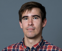
Speaker 1: Eric D. GLOWACKI
Central European Institute of Technology, Chech Republic
Eric Głowacki is a professor of biomedical engineering at the Brno University of Technology in the Czech Republic, and leads the Laboratory for Bioelectronic Materials and Devices at the Central European Institute of Technology, CEITEC, Brno. His background is in physical chemistry (MSc 2009 University of Rochester, USA), (PhD 2013, Johannes Kepler University in Linz, Austria) During postdocs in Linz (2013-2016), his research interest shifted into the field of electrophysiology, bioelectrochemistry, and neuromodulation. In 2016, he moved to set up and independent group at Linköping University in Sweden, within the Wallenberg Center for Molecular Medicine, working on neurostimulation devices. In 2020, he was awarded the ERC Starting Grant, with which he moved his group to CEITEC. His group conducts fundamental research, as well as applied collaborations with companies. The research is dedicated to neural interface technologies and bioelectronic medicine and fundamental exploration in neuromodulation and reactive oxygen species in physiology.
Title:
Electrochemical considerations in bioelectronics interfaces: opportunities and caveats of charge-transfer reactions
Summary:
Studying electric field effects on biological systems, from DC to high AC frequencies, can be confounded by the coexistence charge transfer (CT) reactions. CT redox processes can alter the chemical environment of the biological system being studied. Notable CT reaction effects are: oxygen depletion, pH changes, reactive oxygen species formation, reactive chlorine formation, and electrode corrosion reactions resulting in metal ion release. This tutorial aims to be a practical primer that will overview the common CT reaction types and how to recognize and quantify them. I will cover how CT is affected by frequency/pulse length and by the electrode type. On one hand, I will discuss CT mitigation strategies, on the other hand I will provide some cases where CT can be useful in certain experiments. The notable example is using CT reactions to control oxygen gradients, creating anoxic/hypoxic regions or hyperoxic regions. This concept can be carried further to control reactive oxygen species concentrations. The goal is to provide a better understanding of fundamental electrochemistry in the context of biophysical experiments

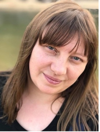
Speaker 2: Anna Guller
Macquarie Medical School, Macquarie University, Australia
Dr Anna Guller has a unique educational and professional background, combining experimental pathology and biophysics, with over 30 years of experience in disease modelling in vivo and in vitro and validation of innovative experimental treatments. Over the last decade, her research has focused on 3D in vitro models of various cancers, extracellular matrix biology, and nanotheranostics. More recently, she has been investigating the response of non-neuronal brain cells to transcranial magnetic stimulation (TMS) and static magnetic fields, exploring their potential effects on glioblastoma and neuroinflammation. As a Senior Scientist at the Children’s Cancer Institute in Australia, she integrates these approaches with emerging therapeutic technologies that leverage physical factors and novel biomaterials to advance translational research.
Title:
3D Cell Cultures and Tissue Engineering in Bioelectromagnetism: Applications and Pathways to Integration
Summary:
Three-dimensional (3D) in vitro cell cultures are the foundation of tissue engineering, enabling the reconstruction of tissues outside the body for research and therapeutic applications. While underutilized in bioelectromagnetism (BioEM) research, 3D cultures offer a more biologically relevant alternative to traditional 2D models and a more ethical, scalable, and cost-effective option than animal testing. The 3D in vitro models provide new opportunities for studying how cells and tissues respond to electric, magnetic, and electromagnetic fields and interact with novel materials sensitive to such fields. This talk will outline practical aspects for generating 3D cultures and detecting the experimental effects, principles of selecting the 3D in vitro models for BioEM purposes, and real-world applications of these platforms. Finally, it will also explore cross-disciplinary integration, highlighting how BioEM can not only benefit from using the 3D cultures but also contribute to advancing extracorporeal tissue reconstruction and preservation, nanotechnology and pharmacological research. These synergies have the potential to improve experimental reproducibility while reducing reliance on animal models, support the transition towards more sustainable research practices, and accelerate the clinical translation of scientific discoveries.

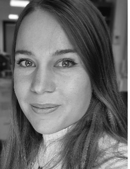
Speaker 3: Leslie Vallet
CNRS, Gustave Roussy, France
Leslie Vallet received the M.Sc. degree in chemical engineering from CPE Lyon Engineering School, Villeurbanne, France, in 2018, associated with one-year exchange study period at the Ecole Polytechnique Fédérale de Lausanne, Lausanne, Switzerland, specializing in biology. She completed her Ph.D. at CNRS – University Paris-Saclay UMR 9018 in Gustave Roussy European Cancer Centre, Villejuif, France, working on research projects involving pulsed electric fields-mediated electroporation with therapeutic applications in oncology and regenerative therapies, as well as the use of dielectrophoresis as a tool for the characterization and cell separation of differentiating mesenchymal stem cells. She is currently working on research projects investigating mechanisms of membrane permeabilization in cells exposed to 18 GHz and THz radiation at the Royal Melbourne Institute of Technology, Melbourne, Australia.
Title:
Dielectrophoresis of mesenchymal stem cells in differentiation: A tool for cell characterization and cell sorting.
Summary:
Mesenchymal stem cells (MSCs) are adult multipotent stem cells naturally able to give rise to different cell types of connective tissue, but also to other specialized cell types under certain conditions. MSCs also exhibit interesting secretory activities, or can even come to the rescue of damaged cells in their environment. Therefore, the use of MSCs in regenerative therapies has attracted considerable interest over the last decades. However, MSC populations exhibit high heterogeneity, which adds complexity to their study in vitro as well as in clinical applications. When transplanted into the body, lack of appropriate cell homing, cell death and rapid clearance, or inappropriate differentiation are limiting factors for the efficiency of MSC-based therapeutic applications. Therefore, the characterization of properties specific to MSCs in differentiation compared to their undifferentiated counterpart, which could lead to the development of non-damaging cell separation methods, would benefit both research and clinical applications. Dielectrophoresis (DEP) is a label-free, rapid processing technique that can be used to characterize the electrical and dielectric properties of the cells and to perform cell electromanipulation without causing cell damage. Using a 3DEP system (LABtech, UK), we evaluated the dielectrophoretic behavior of MSCs during their adipogenic and osteogenic differentiation, acquiring DEP spectra (20 frequencies between 10kHz and 40MHz) at each week of differentiation. Very interestingly, we observed a significant decrease in both membrane permittivity and conductivity in both osteogenic and adipogenic differentiation pathways, as early as in the first week of differentiation, before the appearance of any morphological changes. Later, the evolution of these parameters was less significant. Some other cell properties affecting the dielectrophoretic behavior evolved throughout the differentiation process. In light of these observations, simulations have shown that DEP in combination with a microfluidic channel can be used to perform cell separation of differentiating cells from the first week of the differentiation process. This approach would have the potential to isolate either pure populations of undifferentiated MSCs or populations of pre-differentiated cells devoid of undifferentiated MSCs.